Vet Professionals
Bristol Vet Specialists is one of the most extensive veterinary referral facilities in the country. With a broad range of specialities, cutting-edge equipment, and a top team of specialists.
Pet Owners
Learn about your pet’s journey and find all the information you need to ensure that your pet receives the best possible care at our hospital. You can easily locate our hospital and learn about the various services we offer.
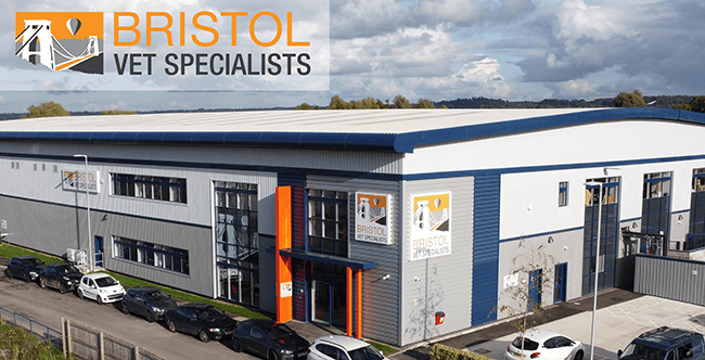
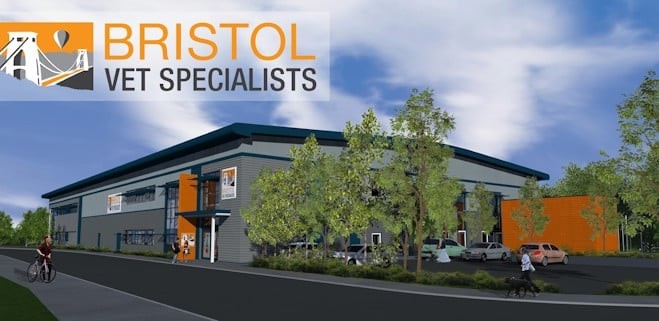
Veterinary Referral Hospital in Bristol
We are one of the largest and most advanced veterinary referral centres in the UK.
Why should you come to us?
At Bristol Vet Specialists we’re proud to provide a diverse range of specialist veterinary referral services. Our services include anaesthesia & analgesia, cardiology, dermatology, diagnostic imaging, emergency and critical care, internal medicine, neurology and neurosurgery, oncology (including radiotherapy), orthopaedic and soft tissue surgery.
With a team of highly trained specialists, advanced equipment, and experienced nursing staff, we’re equipped to handle a wide range of cases from all over the country. Our top priority is providing exceptional care to every patient we see, so you can rest assured that your pet friend is in good hands.
Latest news
£13.5m state-of-the-art Bristol Vet Specialists hospital opens
Largest veterinary hospital in the South West and South WalesEmploying some of the most qualified and experienced veterinary professionals in...
Bristol Vet Specialists is opening October 2023!
We are delighted to confirm that Bristol Veterinary Specialists (BVS) will be welcoming its first patients on Monday 23rd October! Whilst we...
Ophthalmology and Dentistry announced at Bristol Vet Specialists
Bristol Veterinary Specialists, are happy to announce two new clinicians. Kerri-Lee Dobbie, who will head up our ophthalmology referral service, and Jose Carlos Almansa Ruiz, who will be leading our first Dentistry and Maxilliofacial surgery discipline.
CPD Event: CardioVet Symposium; Advancing Cardiology in Veterinary Practice
Join our amazing cardiology team from across CVS Referrals for an informative and interactive CPD day, exploring the latest advancements in...
Here to help
Whether you are a vet phoning for advice, an owner phoning for an update, or if you have any questions, please get in touch and chat to a member of our team who are here to help you.